Caspases and Their Role in Apoptosis: The Cellular Suicide Pathway

Introduction

It is an important physiological process that helps to eliminate unwanted and damaged cells in a structured way through a process called apoptosis, or programmed cell death. This is important in controlling the rate of their death and reproduction and therefore allows organisms to live on. In this process, a group of protease enzymes collectively called caspases is central. These enzymes are critical components in the regulation of the machinery that leads to apoptosis so that the cells are most effectively disassayed. Apoptosis not only has the function of the physiological process that disrupts organisms’ development but also has the function of the pathogenic process protection against diseases, including cancer, where defects in the process lead to uncontrolled cell proliferation. This article also focuses on caspases and their functions in apoptotic processes, regulation and function, and their potential uses in therapeutic utilities.

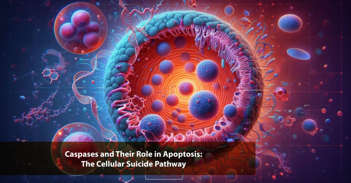
Caspases: Key Players in Apoptosis

Caspases are a group of cysteine proteases that specifically cleave target proteins at aspartyl bonds. These proteases are synthesized as inactive precursors and need to be activated by limited proteolysis. When initiated, caspases set a cellular process in a sequence of events and the disintegration of cellular structures for a defined and timed cell demise. There are two main types of caspases involved in apoptosis: cysteine proteases named initiator caspases, including caspase-8 and caspase-9, along with effector caspases, including caspase-3, caspase-6, and caspase-7.

Initiator caspas are involved in sensing proapoptotic signals and then initiating effector caspas. Effector caspases, therefore, are involved in the proteolytic cleavage of structural and functional proteins within the cell that are characteristic of apoptosis, such as cell shrinkage, chromatin condensation, and DNA fragmentation.

Yearwise Publication Trend on caspases

Find publication trends on relevant topics

Apoptotic Pathways

Apoptosis occurs through two main pathways. It has been classified into two types, namely: the intrinsic, or mitochondrial-dependent pathway, and the extrinsic, or death receptor-mediated pathway. Studies have established that both pathways lead to caspase activation, although the processes of their initiation are diverse.

However, the intrinsic pathway is activated by intracellular signals such as DNA damage, oxidative stress, or the presence of oncogenes. This pathway is well regulated by the proteins that belong to the Bcl-2 family of proteins that regulate the free release of cytochrome c from the mitochondria. When cytochrome c is released to the cytosol, it binds to Apaf-1 and caspase-9 to form a structure known as an apoptosome. The apoptosome in turn activates caspase-9, which activates the effector caspases that result in cell death.

The extrinsic pathway is activated through the binding of pro-apoptotic ligands such as Fas ligand or TNF-α with Fas receptor or TNF receptor, respectively, present on the cell membrane. surface. This interaction brings in adaptor proteins, which include FADD that in turn activate caspase-8. Depending on its cleavage, Caspase-8 can directly activate effector caspases or cleave the pro-apoptotic protein Bid, which in turn results in the release of mitochondrial cytochrome c and the initiation of the intrinsic caspases.

Execution of Apoptosis

After initiator caspases are activated, they cleave and activate effector caspases that are enlisted for deconstructing the cell. Effector caspases target several key cellular components: Effector caspases target several key cellular components:

PARP (Poly ADP-Ribose Polymerase): One of the fundamental proteins involved in the process of repair of DNA. Its slicing nature does not allow any attempt by the cell to heal itself.

Lamins: There are peripheral proteins that have the responsibility of preserving the structural framework of the nuclear envelope. Their degradation leads to nuclear disassembly. Denial of the characteristics results in nuclear disassembly.

Cytoskeletal Proteins: Down-regulation of some proteins, such as actin, experiences proteolytic cleavage resulting in the disassembly of the cytoskeleton and shrinkage of the cell.

These substrates are severed constantly and sequentially so that the cell that comprises the tissue is conveniently eliminated without harming the neighboring tissues.

Recent Publications on caspases

Find publications on relevant topics

Regulation of Caspase Activity

Caspases have been reported to be strictly regulated to avoid scenarios where the cell death process will be instigated accidentally. One contribution to the regulation of peptidergic signaling is their synthesis in an inactive form. They are also controlled by endogenous proteins that suppress the activation of caspases, inclusive of IAP, which snap onto active caspases and neutralize them. For example, XIAP binds with caspase-3 and caspase-9 and thus prevents apoptosis.

Also, the bcl-2 gene product, a group of proteins, is reported to be involved in the regulation of apoptosis through the control of mitochondrial membrane permeability. Bax, a Bak-regulating protein, promotes apoptosis, while Bcl-2 and Bcl-xL inhibit cell death from an intrinsic pathway link.

Caspases in Disease and Therapy

Abnormalities in apoptotic regulation are strongly related to many diseases, most notably cancer. Tumor cells undergo mutation, and there is failure of the body to activate apoptosis, leading to uncontrollable growth of the cancerous cells. Wong et al., 2002 It may, therefore, be characterized by overexpression of anti-apoptotic proteins such as Bcl-2 or loss of pro-apoptotic proteins such as Bax, thereby preventing the induction of apoptosis in cancer cells. Likewise, alterations of caspases or their modulators may interfere with apoptotic pathways, as cancer cells can live through stimuli that in normal circumstances induce cell death.

The therapies directed to modulate apoptosis in cancer cells have received increasing interest. One such strategy is the BH3 mimetics, which are drugs that have been developed to act similarly to the pro-apoptotic Bcl-2 proteins. These drugs are capable of stimulating the intrinsic apoptotic pathway in cancer cells, thus leading to cell death.

Apart from cancer, caspases are involved in other diseases too, as mentioned below. For instance, high levels of apoptosis are involved in the development of neurodegenerative diseases such as Alzheimer’s and Parkinson’s disease. Under these circumstances, the suicide of the neurons through apoptosis and regressive effects on cognition and motor function occur. Knowledge of caspase regulation in these cases provides possible therapeutic strategies for avoiding undesirable cell death associated with neurodegenerative disorders.

In autoimmune diseases, on the contrary, apoptosis failure may result in the survival of auto-reactive immune cells and therefore more chronic inflammatory reactions and tissue destruction. Hence, control of caspase activity could also be useful in autoimmune disease treatment.

Conclusion

Caspases are involved in the apoptosis reaction; they are the initiators and the effectors of regulated cell death. This is through their regulation that ensures that the removal of cells is well done, with the aim of not causing harm to the neighboring tissue. Abnormalities in the regulation of apoptosis and the failure to activate caspase or the overexpression of survival proteins lead to the development of diseases such as cancer. Recent advancements in therapeutic agents have focused on the apoptotic machinery as a potential treatment for cancer, cell death diseases, and auto-immune ailments, as our understanding of caspases continues to expand. The possibility to regulate the activity of caspases is one of the most powerful resources for fighting these diseases and for the development of new types of treatment in the future.

References

  1. Wu, X., Senechal, K., Neshat, M.S., Whang, Y.E. and Sawyers, C.L., 1998. The PTEN/MMAC1 tumor suppressor phosphatase functions as a negative regulator of the phosphoinositide 3-kinase/Akt pathway. Proceedings of the National Academy of Sciences95(26), pp.15587-15591.
  2. Thornberry, N.A. and Lazebnik, Y., 1998. Caspases: enemies within. Science281(5381), pp.1312-1316.
  3. Herrmann, J.L., Beham, A.W., Sarkiss, M., Chiao, P.J., Rands, M.T., Bruckheimer, E.M., Brisbay, S. and McDonnell, T.J., 1997. Bcl-2 suppresses apoptosis resulting from disruption of the NF-κB survival pathway. Experimental cell research237(1), pp.101-109.
  4. Whang, Y.E., Wu, X., Suzuki, H., Reiter, R.E., Tran, C., Vessella, R.L., Said, J.W., Isaacs, W.B. and Sawyers, C.L., 1998. Inactivation of the tumor suppressor PTEN/MMAC1 in advanced human prostate cancer through loss of expression. Proceedings of the National Academy of Sciences95(9), pp.5246-5250.
  5. Maehama, T. and Dixon, J.E., 1998. The tumor suppressor, PTEN/MMAC1, dephosphorylates the lipid second messenger, phosphatidylinositol 3, 4, 5-trisphosphate. Journal of Biological Chemistry273(22), pp.13375-13378.
  6. Furnari, F.B., Lin, H., Huang, H.J.S. and Cavenee, W.K., 1997. Growth suppression of glioma cells by PTEN requires a functional phosphatase catalytic domain. Proceedings of the National Academy of Sciences94(23), pp.12479-12484.
  7. Li, J., Yen, C., Liaw, D., Podsypanina, K., Bose, S., Wang, S.I., Puc, J., Miliaresis, C., Rodgers, L., McCombie, R. and Bigner, S.H., 1997. PTEN, a putative protein tyrosine phosphatase gene mutated in human brain, breast, and prostate cancer. science275(5308), pp.1943-1947.
  8. Herrmann, J.L., Beham, A.W., Sarkiss, M., Chiao, P.J., Rands, M.T., Bruckheimer, E.M., Brisbay, S. and McDonnell, T.J., 1997. Bcl-2 suppresses apoptosis resulting from disruption of the NF-κB survival pathway. Experimental cell research237(1), pp.101-109.

Top Experts on “caspases